All you need is a good history and physical, and to follow the signs described. Very rarely will you need an expensive diagnostic workup like CT scans, MRIs, ultrasounds, endoscopies and laparoscopies.
The diagnosis is fairly straightforward and initially suspected within 25 to 40 minutes, primarily on history and clinical exam. Awareness and strong suspicion of this condition is helpful. A careful history may reveal:
- Pain that gets worse with physical activities.
- Previous history of lower abdominal surgeries, especially multiple surgeries.
- History of enthesopathies (tendonitis) in other parts of the body, such as tennis elbow or plantar fasciitis.
- Very few gastrointestinal symptoms.
The physical exam
On physical exam, careful assessment of the bony points, including the pubic bone and ASIS (anterior superior iliac spine), is critical in suspecting the syndrome. Tenderness was most common at 3 locations on the pubic bone: over the pubic tubercle laterally (98%), over the pubic crest at the midline (60 to 70%) and over the inferior ramus of the pubic bone (40 to 50%). The majority of these patients had radiating pain over the medial aspect of the thigh, all the way to the knee. Nearly 40% of all patients had marked tenderness over the insertion of the adductor longus tendon to the pubic bone (positive adductor sign).
Detailed examination of the abdomen, pelvis (including vaginal and rectal exam), genitals, hip joints, lower back, coccyx, ischial tuberosities and thighs is important in arriving at the diagnosis.
The omission of the exam of the pubic bone and anterior superior iliac spine on physical exam of the abdomen, specifically in patients with lower abdominal pain, is the most embarrassing cause of a missed diagnosis.
Wasu’s Triad and Triad Plus
In females
Wasu’s Triad
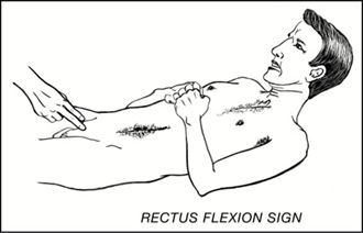
Rectus flexion sign
The examiner’s hand is placed on the insertion of the rectus muscle over the pubic bone. If the patient experiences severe pain on flexion of the rectus muscle when attempting to sit up, it is considered a positive sign.
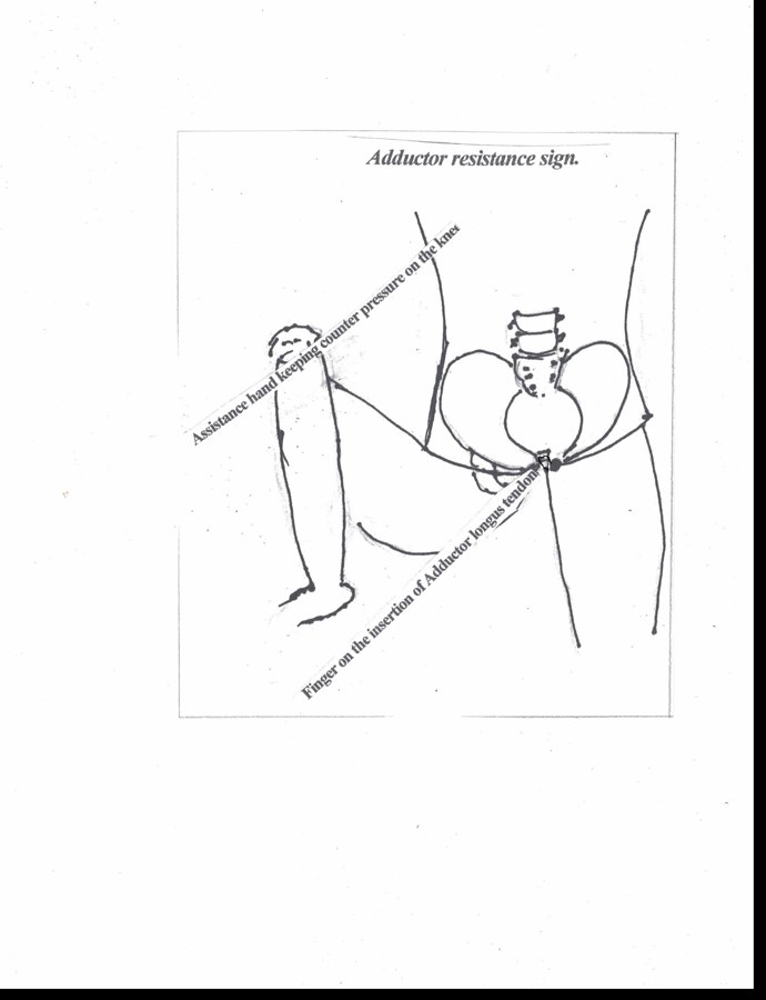
Adductor resistance sign
Positive if the patient experiences pain when the examiner places a finger over the insertion of the adductor longus tendon and asks the patient to adduct the bent knee against the assistance hand.
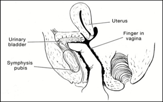
Vaginal hook sign
Positive if the patient experiences pain over the suprapubic area on vaginal exam when hooking fingers anteriorly over the pubic bones.
In males
Wasu’s Triad Plus
Rectus flexion sign
As in females: severe pain over the pubic bone when the rectus muscle is flexed while attempting to sit up.
Adductor resistance sign
Pain at the insertion of the adductor longus tendon on resisted adduction of the bent knee.
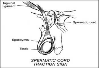
Spermatic cord traction sign
Examined in a standing position: traction of the spermatic cord produces radiating pain over the pubic bone. The spermatic cord itself is not tender when examined lying down.
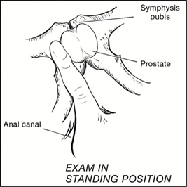
Paraprostatic sign
On digital rectal exam there is mild or no pain over the prostate gland itself, but tenderness and muscle spasm lateral to the prostate. This suggests chronic pelvic pain, also known as nonbacterial prostatitis.
Diagnostic studies
Different authors have used several diagnostic studies to confirm the diagnosis of this condition, such as CT scan, bone scans, ultrasound, and plain x-rays. Some authors have found MRI useful to detect bone marrow edema, tear or degeneration of the tendons, etc. The value of herniography is controversial. Kluin et al. have used diagnostic laparoscopy to confirm the diagnosis in 10 out of 14 athletes.
Final confirmation is conducted by administering a series of steroid injections and observing the patient’s response.